
I understand the body best when I build it up from the smallest to the largest. First, I look at cells. Then I show how tissue types develop from them and why their structure matters. I also explain how several tissues form an organ and how organ systems work together. This way, you’ll understand what epithelium protects, connective tissue supports, muscle moves, and nerve tissue controls. Additionally, I clearly distinguish between parenchyma and stroma so you can classify their functions. This helps you learn terms faster.
1. Why Tissues Are More Than Just Clusters of Cells
When I want to understand the body, I don’t start with large organs. I start with cells. However, cells rarely work alone. They almost always work as a team. This is precisely where the topic of tissue types begins.
I understand a tissue as a functional unit. It consists of many cells that are similarly specialized. Furthermore, these cells are arranged in a clear structure. This structure plays a crucial role in determining performance. Therefore, it’s not enough to only know individual cells. I also need to know how they connect, how they communicate, and how they work together to solve problems.
At the same time, when I think of tissue, I don’t just think of “material.” I think of processes. One tissue protects. Another moves. Yet another conducts signals. This is how a reliable overall function emerges from many small parts.
One point is particularly important to me here. A tissue doesn’t “become the way it is” by chance. Cells adapt to their job. They change their structure. They change their shape. They change their connections to neighboring cells. Thus, biology becomes a system that I can logically explain.
2. From Micro to Macro: How the Body Builds Itself
I recognize a clear hierarchy within the body. It helps me to easily organize complex information. First comes the cell. Then comes the cell cluster. From this, tissue develops. Several tissues form an organ. Several organs work together as an organ system. Finally, the sum of these makes up the whole person.
This sequence sounds simple. Yet it resolves many misconceptions. Because I can often only explain an organ’s function if I understand the corresponding tissue. For example, I can’t meaningfully discuss kidney function if I don’t understand which cell types filter, transport, and seal there. Similarly, I can’t accurately categorize movements if I only see muscle tissue as “muscle.”
Furthermore, the hierarchy helps me in practice. When symptoms occur, I ask myself: Where is the problem likely to lie? Is it in the cell? Is it in the tissue cluster? Or is it in the coordination of several organs? This allows me to quickly gain structure to medical issues.
I also notice: The higher I go in the hierarchy, the stronger the cooperation becomes. An organ system never solves tasks alone. It always works in a network. That’s why a clear understanding of its structure is so valuable.
3. Cell Specialization Explained Simply: Organelles as Toolboxes
I see each cell as a tiny workshop. In this workshop lie tools. These tools are called cell organelles. Depending on the task, the cell prioritizes different tools. This is why we speak of specialization.
Initially, specialization doesn’t arise from a desire, but from a need. A cell receives signals from its environment. It receives developmental programs. It then aligns its production accordingly. It expands certain structures more extensively. It uses others less. This makes it better suited to its job.
An example makes this tangible. When I think of immune cells, I see active “clean-up crews.” These cells ingest pathogens. Then they break down the ingested components. For this, they need many small breakdown units inside them. These units contain enzymes. These enzymes break down proteins and other substances. In this way, the cell can neutralize harmful particles. At the same time, it protects the surrounding tissue.
However, specialization also comes at a price. A highly specialized cell is usually less flexible. It invests energy in its primary function. Therefore, it is less able to perform other tasks. Furthermore, some specialized cells rarely or never divide. This influences regeneration.
Nevertheless, specialization is crucial. Without it, there would be no clear tissue types. There would only be “average cells.” The body would then perform many tasks too slowly or too imprecisely. That’s why I always base my understanding on this principle.
4. The Four Basic Tissues at a Glance
When I classify tissue types, I use four basic groups. These groups appear throughout the body. They differ in structure, material, and function. At the same time, they complement each other.
First, there is epithelial tissue. I use this term to describe surfaces and linings. It defines boundaries. It protects. It exchanges substances. It also often controls what is allowed in and what must stay out.
Second, there is connective and supporting tissue. I see it as a framework, cushion, and connecting layer. It holds organs in shape. It anchors structures. It also forms the environment for blood vessels and nerve pathways. Depending on the variant, it can appear soft, firm, or even hard.
Third, there is muscle tissue. I associate it with the topic of movement. This applies to visible movements. But it also applies to internal movements, for example, in hollow organs. Therefore, muscle tissue is not just about “exercise.” It is also about transport and control.
Fourthly, there is nerve tissue. I connect it with information. It receives stimuli. It processes stimuli. It transmits commands. In doing so, it coordinates many functions that would otherwise be incompatible.
I use this classification as a compass. From there, I can delve deeper. I can then clearly describe each tissue without getting lost in the details.
I use this classification as a compass. From there, I can delve deeper. I can then clearly describe each tissue without getting lost in the details.
5. Epithelial Tissue: The Barrier That Also Controls
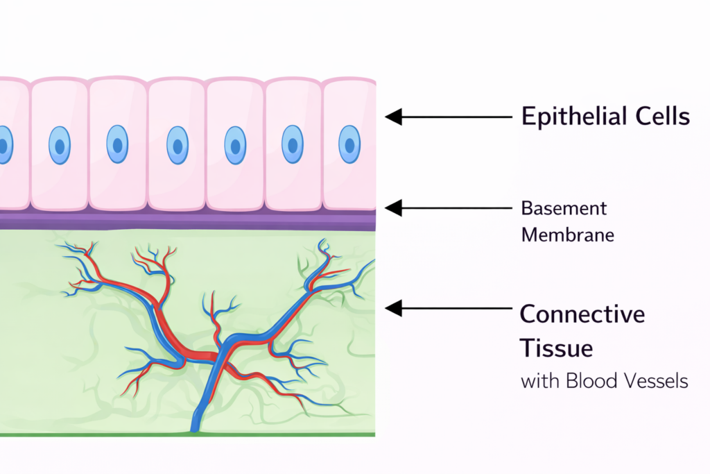
When I think of epithelial tissue, I first think of boundaries. Epithelium covers the body’s surface. It also lines internal cavities. This creates a controlled interface with the outside world and the inside.
For epithelium to provide reliable protection, it must function tightly. I imagine the cells like closely spaced paving stones. Gaps must not remain open. Otherwise, the body loses too much water, or substances penetrate uncontrollably. Therefore, epithelial cells use strong cell contacts. These contacts connect cell to cell, creating a closed layer.
At the same time, epithelium must remain flexible. Some epithelia protect against friction. Others absorb substances. Still others produce secretions, such as mucus. This is why epithelia vary greatly in shape and thickness. Nevertheless, the basic principle remains the same. I see a densely organized layer of cells with a clear orientation.
Another point is crucial. Epithelium generally does not have its own blood vessels. This may sound surprising at first. Nevertheless, the supply of nutrients and oxygen functions. Because the tissue beneath it carries blood vessels right up to the boundary. From there, oxygen and nutrients continue their journey. They travel through tissue fluid. They cross a thin separating layer. Then they reach the epithelial cells.
I call this separating layer the basement membrane. I use it as a clear boundary between epithelium and connective tissue. At the same time, it acts like a foundation. It holds the epithelial cells in place. It also influences how cells grow and regenerate.
That’s why I summarize epithelium in this way. It forms a dense covering. It protects and regulates. It works with the underlying connective tissue as a supply partner. This creates a surface that remains both robust and functional.
6. Connective and Supporting Tissue: The Body’s Framework with Extra Matrix
Connective and supporting tissue is everywhere. I find it between organs. I find it in organs. I find it as a covering, as padding, and as a framework. That’s why it’s one of the most common types of tissue.
I see three core ideas. First, it connects structures. Secondly, it creates space and allows for movement between organs. Thirdly, it provides support. This is precisely why we often speak of the “stroma” when referring to the supporting framework of an organ.
Its structure differs significantly from epithelium. Here, it’s not just the cell that matters. The environment between the cells is also crucial. This environment is called the extracellular matrix. I conceptually divide it into two parts: ground substance and fibers.
The ground substance consists largely of water and gel-like components. It acts like a finely tuned environment. It can be very fluid, or it can be gel-like. In some tissues, it even becomes solid. This occurs particularly where the body needs stability.
The fibers give the matrix direction and resilience. I primarily distinguish between two fiber families. Collagen fibers provide tensile strength. Elastic fibers provide extensibility and resilience. Depending on the ratio of fibers, the tissue feels firm or soft.
Now I come to the cells. I distinguish two roles for them. Some cells reside permanently within the tissue. Others migrate in and out.
I see the stationary cells as a construction and maintenance team. Fibroblasts produce new matrix. They build fibers and ground substance. If the tissue remains stable, more mature forms like fibrocytes tend to dominate. They maintain the structure. At the same time, they react to stress, injury, or remodeling.
I see the migrating cells as a safety and repair team. This includes various immune cells. They appear when inflammation develops. They clean up. They coordinate reactions. This is why connective tissue often becomes the site where defense processes become visible.
Furthermore, there are several types of connective and supporting tissue. I classify them according to function. Adipose tissue stores energy and provides cushioning. Loose connective tissue allows for mobility. Dense connective tissue supports and anchors. Reticular tissue forms fine networks, for example, as the framework in certain organs. Cartilage supports and cushions. Bone bears loads and protects because it is mineralized.
Therefore, for me, connective and supporting tissue is more than just “filler.” It’s the infrastructure. It connects everything. It supplies pathways for blood vessels and nerves. It stabilizes organs. And it reacts very actively to stress.
7. Muscle Tissue: The Engine in Three Variants
Muscle tissue represents movement. However, by movement I don’t just mean sports. I also mean internal transport. I also mean the work of pumps, valves, and sphincters. That’s why I always look at muscle tissue in its various forms.
I first distinguish between smooth muscle and striated skeletal muscle. Additionally, there is cardiac muscle tissue as a special form. Each variant solves a different problem.
Smooth muscle works without conscious control. I find it in the walls of hollow organs, such as the stomach and intestines, as well as parts of the bile ducts and many blood vessels. This muscle moves contents along. It mixes. It regulates diameter. It works rather slowly, but reliably. Furthermore, it often exhibits rhythmic activity.

An important point is its control mechanism. Smooth muscle doesn’t always wait for commands from the brain. It can utilize local signals. Networks of nerve cells in the organ wall provide the pace and direction. Receptors also signal when the contents change, generating local impulses. These impulses trigger contraction. That’s why the transport remains stable even when I’m not consciously thinking about it.
Striated skeletal muscle works differently. I use it for quick, targeted movements. I raise my arm. I walk. I maintain my posture. Here, I make conscious decisions. Therefore, the command originates from the central nervous system. Furthermore, this muscle works quickly. It works powerfully. However, it doesn’t automatically follow its own rhythm. It reacts to clear signals.

Cardiac muscle tissue exists between these two worlds. It works involuntarily. At the same time, it works in a highly organized and continuous manner. It combines force with rhythm. It utilizes its own pacemaker structures. Nevertheless, it reacts to signals from the nervous system and to hormones. This allows it to adjust its performance to the demand.
When I compare muscle tissue, I always consider four questions: Am I working voluntarily or involuntarily? Am I working quickly or slowly? Am I working rhythmically or freely? And where does the impulse originate? With these questions, I can accurately classify every muscle activity. This also helps me understand better why the body “moves” differently internally than externally.
8. Nerve Tissue: Communication, Processing, Control
If I view the body as a system, then nerve tissue is both the control center and the network of connections. It links sensory impressions with decisions. It connects decisions with actions. It also connects organs to each other so that everything stays in sync.
I divide nerve tissue into two main groups. I distinguish between nerve cells and glial cells. Both belong together. Nevertheless, they fulfill different tasks.
8.1 Neurons and Glia: Clear Roles, Clear Logic
Nerve cells are also called neurons. They transmit excitation. They receive stimuli. They process information. And they transmit signals to other cells. This creates chains of stimulus, processing, and response.
Glia cells form the supporting environment. I see them as a service team. They support nerve cells. They protect them. They insulate the pathways. They also provide nerve cells with essential conditions so that they function stably.
Therefore, for me, the following applies: Without neurons, there would be no signal transmission. Without glia, there would be no stable environment for this signal transmission.
8.2 Why some cells no longer divide
I keep one principle in mind. Many nerve cells hardly divide anymore. They are extremely specialized. They invest energy in function. As a result, they often lose the ability to divide. This explains why damage to the nervous system is sometimes more difficult to repair.
Glial cells more frequently retain this ability. Therefore, they can react, remodel, and provide support in many situations. This keeps the tissue functional, even when stressed.
8.3 Structure of a Neuron: Direction Decides

To truly understand nerve cells, I look at their structure. A neuron consists of a cell body and extensions. I distinguish between three terms here.
The cell body is called the soma. The nucleus is located there. Many metabolic processes take place there. The cell processes a large portion of its signals there.
Then come the dendrites. I think of them as receiving arms. They often branch out. They collect signals from the surrounding area. Then they transmit these signals to the soma.
The axon is the outgoing extension. It carries signals away from the soma. It can be very short. But it can also be very long. At its end, it branches out and forms connections with other cells.
This establishes a clear direction. Dendrites lead to the soma. The axon leads away from the soma. This orientation helps me to logically explain nerve impulse conduction.
8.4 Synapses: This is where the switching occurs

At the end of the axon are synapses. These contact points are called synapses. A synapse connects one nerve cell to another cell. This other cell can be another nerve cell, but it can also be a muscle cell or a gland cell.
That’s precisely why I see synapses as switching points. Here, it’s decided whether a signal continues its travel. Here, it’s also decided how strong the effect will be.
8.5 From Electrical to Chemical and Back
I understand the synapse as a translator. An electrical signal travels along the axon. At the synapse, this is often converted into a chemical signal. Then, an electrical impulse or another reaction occurs in the target cell.
In many synapses, there are small vesicles at the end of the axon. These vesicles store messenger substances. These messenger substances are called neurotransmitters. When an electrical signal arrives, these vesicles fuse with the membrane. They then release the messenger substance into a tiny space. This space is called the synaptic cleft.
The messenger substance travels to the other side. The postsynaptic membrane is located there. Matching receptors bind the neurotransmitter there. This triggers a response in the target cell.
This response can take different forms. If the target cell is a neuron, a new electrical impulse is often generated. If the target cell is a muscle fiber, contraction begins. If the target cell is a gland, secretion increases or decreases.
This is how control is established. This is how coordination is established. And this is how behavior, movement, or regulation ultimately arises.
9. Understanding Organs: Who Works, Who Supports
When I talk about organs, I don’t just think about shape and location. I think about structure. An organ almost never consists of just one type of tissue. It combines several tissues because it has to perform multiple tasks simultaneously.
To quickly understand organ structure, I use two terms. I distinguish between parenchyma and stroma. This distinction provides structure.
9.1 Parenchyma: The Team for the Main Tasks
The parenchyma comprises the cells that perform the core functions. These cells determine the typical function of the organ. They provide the performance I associate with the organ.
When I think of the heart, I see cells that contract. These cells generate pumping power. When I think of the lungs, I see structures that exchange gases. These areas ensure the uptake of oxygen and the release of carbon dioxide.
That’s why, for me, parenchyma is the actual working surface of an organ.
9.2 Stroma: The framework, the supply, the pathways
The stroma forms the supporting environment. It often consists of connective tissue. It keeps the organ in shape. It protects sensitive working areas. It also carries blood vessels and nerves into the organ.
These pathways are crucial. Because the parenchyma needs oxygen and nutrients. It also needs removal. Furthermore, it needs regulation. That’s why many pathways converge in the stroma.
I also remember: Many inflammatory processes take place in the stroma. There, immune cells encounter blood vessels. Swelling and remodeling occur. This often alters the function of the entire organ.
10. Organ Systems in the Human Body: The Major Functional Networks
An organ rarely works in isolation. Therefore, I classify organs into organ systems. An organ system groups together organs that perform a common primary function. At the same time, each organ remains a specialist. This creates a network of specialists.
Here, I list the most important systems that I constantly use in everyday life and in my studies.
10.1 Musculoskeletal System
This includes bones, joints, ligaments, and muscles. This system enables movement. It also provides stability and protection. Furthermore, bones store important minerals.
10.2 Digestive System
This system ingests food. It breaks down nutrients. It absorbs and distributes them. This also includes organs that produce enzymes and bile.
10.3 Respiratory System
This system brings oxygen into the body. It removes carbon dioxide. It works closely with the circulatory and nervous systems.
10.4 Cardiovascular System
The heart pumps blood. Vessels transport it. This is how oxygen and nutrients reach the cells. At the same time, the circulatory system carries away metabolic waste products.
10.5 Blood and Blood-Forming Organs
Here, I’m thinking of blood as a transport and defense medium. I’m also thinking of organs that produce and break down blood cells. This keeps the system renewable and adaptable.
10.6 Lymphatic System
This system collects tissue fluid. It transports it back into the blood. It is also central to defense and filtration.
10.7 Endocrine System
This is about hormones. Hormones regulate long-term processes. They influence growth, metabolism, stress responses, and reproduction. Therefore, this system works slowly but very powerfully.
10.8 Urogenital System
This system connects excretion and reproduction. The urinary organs regulate water, salts, and many substances in the blood. The reproductive organs ensure continuation and hormone production.
10.9 Skin and Skin Appendages
The skin protects. It regulates temperature. It prevents water loss. Furthermore, it is a sensory organ. Hair, nails, and glands are part of its appendages.
10.10 Sensory Organs
These organs receive stimuli. They translate the outside world into signals. The nervous system then takes over the processing.
10.11 Nervous System
Here I classify the brain, spinal cord, and peripheral nerves. This system coordinates rapid reactions. It connects perception with movement and regulation.
10.12 Immune System
I see this system as a protective net. It recognizes foreign substances. It removes danger. It builds memory. At the same time, it must remain tolerant so that it does not attack the body.
11. How it all works together: three mini-scenarios from everyday life
I understand tissues, organs, and systems best through situations. That’s why I use small scenarios. They show me how cooperation works.
11.1 After eating: Transport, movement, control
When I eat, absorption begins first. This is followed by transport. Smooth muscle moves the contents along. At the same time, glands produce digestive juices. The epithelium absorbs substances. The nervous system coordinates reflexes. Hormones also adjust digestion.
This creates a process that functions without conscious control. Yet it remains highly precise.
11.2 Sprinting: Muscle work, circulation, signaling pathways
When I sprint, I want immediate performance. The nervous system sends rapid commands to skeletal muscles. The muscles contract powerfully. At the same time, the heart increases its pumping capacity. Breathing becomes deeper and faster. Energy reserves are also mobilized.
This shows me: Movement is never just muscle work. Movement is always systemic work.
11.3 Infection: Defense, Tissue Reaction, Supply
When pathogens invade, the local tissue often reacts first. The epithelium tries to block them. If that’s not enough, the immune system kicks in. Connective tissue plays a major role here because many immune cells arrive there. Blood vessels dilate. Fluid leaks out, causing swelling. At the same time, cells migrate in and fight the pathogens.
This explains inflammation as a coordinated reaction, not as a “mistake.” Nevertheless, it can cause problems if it is too severe or lasts too long.
12. Learning and Exam Boost: Typical Pitfalls and My Solutions
I keep seeing the same misunderstandings. That’s why I deliberately collect them. This helps me learn faster and more effectively.
12.1 Tissue, Organ, Organ System: Clearly Distinguishing the Levels
I first ask myself: Am I talking about cells? Am I talking about tissues? Or am I talking about organs? Many mistakes occur because terms are used interchangeably. That’s why I maintain consistency in my understanding.
12.2 Epithelium without blood vessels: still supplied
Many people stumble over the issue of supply. I remember: epithelium is dense and avascular. The underlying connective tissue provides the supply. Diffusion takes care of the rest. This way, protection is maintained, and nutrition still works.
12.3 Parenchyma and stroma: don’t confuse them
I remember a simple rule: parenchyma does the work. Stroma supports and supplies. When I apply this rule, I quickly classify many examples correctly.
12.4 Muscle types: control first, not location
Many people think about location first. I think about control first. Voluntary muscle means skeletal muscle. Involuntary muscle means smooth muscle or heart muscle. Then I look at speed and rhythm.
12.5 Nerve cell and glia: don’t lump them together
I separate function. Neuron conducts and processes. Glia stabilizes and protects. This keeps the terminology clear.
13. Conclusion: The Body as a Modular System from Cell to Organ System
If I truly understand the body, I think in terms of levels. Cells form tissues. Tissues build organs. Organs form organ systems. Ultimately, this results in a functioning organism.
Furthermore, I recognize that each level performs its own tasks. Nevertheless, everything is interconnected. That’s why it’s worthwhile to look at tissue types. It makes organ functions logical. It makes symptoms understandable. And it makes learning easier.
14. FAQ: Frequently Asked Questions about Tissue Types and Organ Structure
What is the most important difference between tissue and organ?
A tissue consists of similar cells with a common task. An organ combines several tissues for a complex function.
Why does the body need four basic tissues?
Because it has to solve four fundamental problems. It needs barriers. It needs stability. It needs movement. And it needs information.
Why is inflammation often visible in the stroma?
Because blood vessels run there and immune cells migrate into it. Signals, cells, and supplies converge there.
Why does smooth muscle work rhythmically?
Because local control centers and receptors in the organ wall trigger activity. This ensures reliable transport, even without conscious control.